Cranial Nerve 7 Nucleus - Cranial nerves & Cranial nerve nuclei : - PPT Powerpoint / There are many cranial nerve mnemonics that can be memorable and rude/lewd.
Cranial Nerve 7 Nucleus - Cranial nerves & Cranial nerve nuclei : - PPT Powerpoint / There are many cranial nerve mnemonics that can be memorable and rude/lewd.. Also, these nerves connect with many of the same brain stem nuclei (dorsal motor nucleus of the vagus, nucleus ambiguus, nucleus solitarius, spinal nucleus of the trigeminal). (the olfactory and optic centers are not represented.) latin nucleus nervi cranialis gray s. Some of each kind are related to. The weber test lateralizes to the right. Because the cranial nerve nuclei are dispersed throughout the brainstem rather than being clustered together, the oral structures may be selectively impaired and should be carefully evaluated.
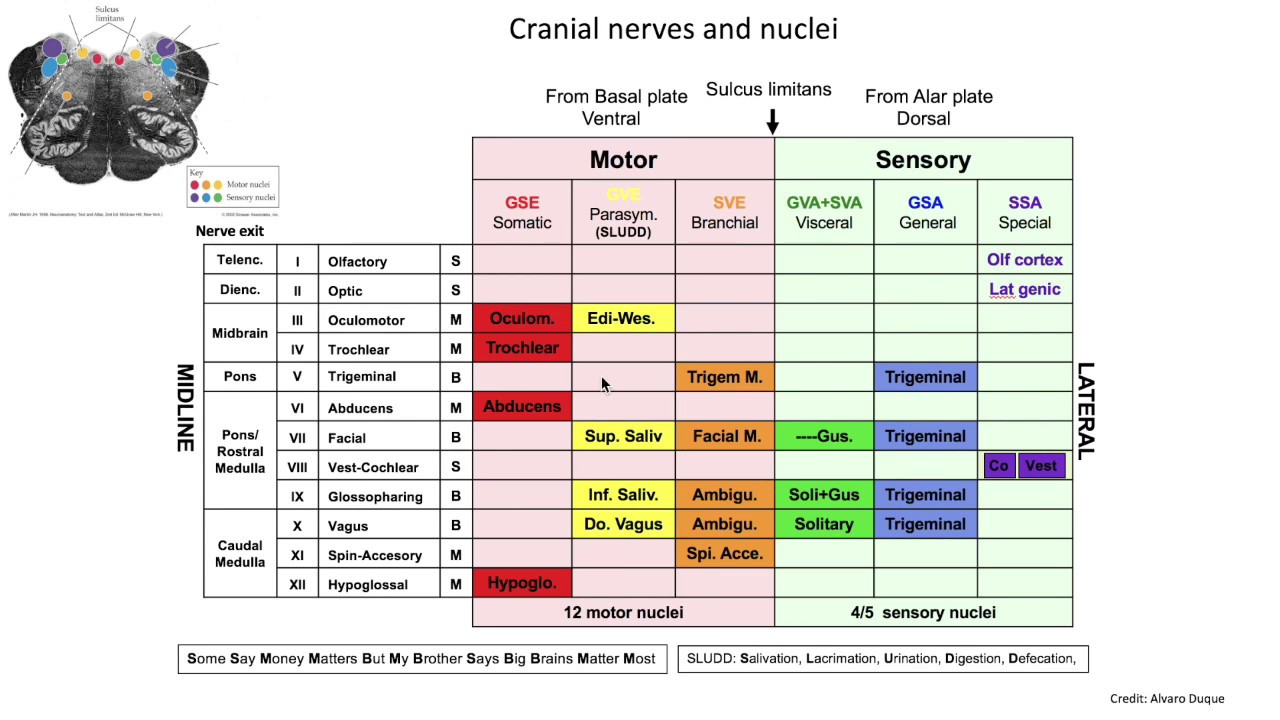
The weber test lateralizes to the right. Cranial nerve nuclei have a generally predictable arrangement. Either way, they can be helpful for remembering the names of the twelve cranial nerves, as well as remembering which nerves are sensory, motor, or both. Taste receptor cells are innervated by one of three cranial nerves (vii, ix, or x), which project topographically into the rostral portion of nucleus of the solitary. The lower cranial nerves are involved in pharyngeal and laryngeal function as well as in movements of the neck and tongue.
Cranial Nerve Nuclei from www.brainkart.com However, the other cranial convey somatic sensory innervation from the skin the position and number of nuclei for each cranial nerve (excluding those serving the special senses) can be worked out using a few basic rules. These are paired on the left and right side of. Locations of cranial nerve nuclei are shown in transverse sections (left), a dorsal view of the brain stem (lower right), and a medial view of the right half somatic efferent nuclei are dark blue and light blue (the latter nuclei innervate pharyngeal arch derivatives). These nuclei are shown schematically in the lower half of fig. The sulcus limitans intervenes between motor and sensory cranial nerve nuclei have a generally predictable arrangement. Cranial nerves relay information between the brain and parts of the body, primarily to and from regions of the head and neck. Neuralation begins at the trilaminar embryo with formation of the notochord within the mesoderm that underlies the ectoderm and do not physically contribute to the nervous system, but is involved with patterning its initial formation. Some of each kind are related to.
Spinal nerves contain sensory and motor fibers.
Cranial nerve nuclei have a generally predictable arrangement. Either way, they can be helpful for remembering the names of the twelve cranial nerves, as well as remembering which nerves are sensory, motor, or both. It supplies motor, sensory and parasympathetic innervation to various structures of the head and neck. Because the cranial nerve nuclei are dispersed throughout the brainstem rather than being clustered together, the oral structures may be selectively impaired and should be carefully evaluated. Cranial nerves are the nerves that emerge directly from the brain (including the brainstem), of which there are conventionally considered twelve pairs. The 7th cranial nerve is mixed nerve containing both sensory and motor components. A cranial nerve nucleus is a collection of neurons (gray matter) in the brain stem that is associated with one or more of the cranial nerves. Some cranial nerves deal specifically with special senses as we have seen. Muscle tone in lower motor neuron damage is flaccid or hypotonic. Cranial nerves relay information between the brain and parts of the body, primarily to and from regions of the head and neck. Also, these nerves connect with many of the same brain stem nuclei (dorsal motor nucleus of the vagus, nucleus ambiguus, nucleus solitarius, spinal nucleus of the trigeminal). Some nuclei retain their original positions in relation to the floor of the fourth ventricle, but some others in the description that follows the nuclei of the third to twelfth cranial nerves are considered as they are located in the brainstem. Cranial nerve nucleus the cranial nerve nuclei schematically represented;
Locations of cranial nerve nuclei are shown in transverse sections (left), a dorsal view of the brain stem (lower right), and a medial view of the right half somatic efferent nuclei are dark blue and light blue (the latter nuclei innervate pharyngeal arch derivatives). Just as the spinal cord receives sensory information from the body surface and deeper tissues and sends motor axons to striated muscles and to the autonomic. Muscle tone in lower motor neuron damage is flaccid or hypotonic. Some of each kind are related to. Some cranial nerves deal specifically with special senses as we have seen.
Cranial Nerve 6: Abducens Nerve from i0.wp.com Locations of cranial nerve nuclei are shown in transverse sections (left), a dorsal view of the brain stem (lower right), and a medial view of the right half somatic efferent nuclei are dark blue and light blue (the latter nuclei innervate pharyngeal arch derivatives). Cranial nerve nucleus the cranial nerve nuclei schematically represented; It supplies motor, sensory and parasympathetic innervation to various structures of the head and neck. The visceral afferent nucleus is orange. Cranial nerve nuclei have a generally predictable arrangement. Cranial nerve palsies can be congenital or acquired. The lower cranial nerves are involved in pharyngeal and laryngeal function as well as in movements of the neck and tongue. These are paired on the left and right side of.
Related online courses on physioplus.
It supplies motor, sensory and parasympathetic innervation to various structures of the head and neck. Just as the spinal cord receives sensory information from the body surface and deeper tissues and sends motor axons to striated muscles and to the autonomic. Cranial nerve seven (cn vii) is responsible for both efferent and afferent modalities in the head and neck including the parasympathetic fibers of cn vii originate in the superior salivary nucleus of the pons and leave the cerebellopontine angle as the nervus intermedius (of wrisberg). Cranial nerves are the nerves that emerge directly from the brain (including the brainstem), of which there are conventionally considered twelve pairs. Cranial nerve palsies can be congenital or acquired. After an introductory section surveying cranial nerve organization and tricky basics such as ganglia, nuclei and brain stem pathways, the nerves are considered in functional groups: Also, these nerves connect with many of the same brain stem nuclei (dorsal motor nucleus of the vagus, nucleus ambiguus, nucleus solitarius, spinal nucleus of the trigeminal). Cranial nerves relay information between the brain and parts of the body, primarily to and from regions of the head and neck. Some cranial nerves deal specifically with special senses as we have seen. Neuralation begins at the trilaminar embryo with formation of the notochord within the mesoderm that underlies the ectoderm and do not physically contribute to the nervous system, but is involved with patterning its initial formation. Either way, they can be helpful for remembering the names of the twelve cranial nerves, as well as remembering which nerves are sensory, motor, or both. Muscle tone in lower motor neuron damage is flaccid or hypotonic. Animal physiotherapy foundation programme online course:
Cranial nerve nuclei have a generally predictable arrangement. The cranial nerve nuclei are made up of the neurons in the brainstem that receive primary sensory inputs or that give rise to motor outputs. Some nuclei retain their original positions in relation to the floor of the fourth ventricle, but some others in the description that follows the nuclei of the third to twelfth cranial nerves are considered as they are located in the brainstem. There are many cranial nerve mnemonics that can be memorable and rude/lewd. Cranial nerves relay information between the brain and parts of the body, primarily to and from regions of the head and neck.
Brain-Splaining EP1: Exploring the Cranial Nerve Nuclei ... from i.ytimg.com Animal physiotherapy foundation programme online course: Cranial nerve nucleus the cranial nerve nuclei schematically represented; Want to learn more about it? Multiple cranial neuropathies are commonly seen in lesions caused by tumors, trauma, ischemia lesions of the trigeminal nerve nuclei (depending on the nuclei affected): The cranial nerve nuclei are made up of the neurons in the brainstem that receive primary sensory inputs or that give rise to motor outputs. (the olfactory and optic centers are not represented.) latin nucleus nervi cranialis gray s. Ipsilateral weakness of muscles of mastication and/or ipsilateral loss of sensation. The sulcus limitans intervenes between motor and sensory cranial nerve nuclei have a generally predictable arrangement.
Cranial nerve palsies can be congenital or acquired.
These are paired on the left and right side of. These nuclei are shown schematically in the lower half of fig. The 7th cranial nerve is mixed nerve containing both sensory and motor components. The lower cranial nerves are involved in pharyngeal and laryngeal function as well as in movements of the neck and tongue. There are many cranial nerve mnemonics that can be memorable and rude/lewd. Cranial nerve palsies can be congenital or acquired. Animal physiotherapy foundation programme online course: Some cranial nerves deal specifically with special senses as we have seen. Spinal nerves contain sensory and motor fibers. Want to learn more about it? Multiple cranial neuropathies are commonly seen in lesions caused by tumors, trauma, ischemia lesions of the trigeminal nerve nuclei (depending on the nuclei affected): Cranial nerves may be affected singly or in groups and knowledge of which nerves are involved helps locate the lesion. The seventh cranial nerve is responsible for eyelid closure during the blink reflex.
You have just read the article entitled Cranial Nerve 7 Nucleus - Cranial nerves & Cranial nerve nuclei : - PPT Powerpoint / There are many cranial nerve mnemonics that can be memorable and rude/lewd.. You can also bookmark this page with the URL : https://sak-laop.blogspot.com/2021/07/cranial-nerve-7-nucleus-cranial-nerves.html
Share Awesome
Belum ada Komentar untuk "Cranial Nerve 7 Nucleus - Cranial nerves & Cranial nerve nuclei : - PPT Powerpoint / There are many cranial nerve mnemonics that can be memorable and rude/lewd."


Belum ada Komentar untuk "Cranial Nerve 7 Nucleus - Cranial nerves & Cranial nerve nuclei : - PPT Powerpoint / There are many cranial nerve mnemonics that can be memorable and rude/lewd."
Posting Komentar